
Oral mucocele is a common lesion of the oral mucosa that results from an alteration of minor salivary glands due to mucous accumulation
Table of Contents
General features of Oral Mucocele:
- Oral Mucocele is a common lesion of the oral mucosa that results from an alteration of minor salivary glands due to mucous accumulation.
- Other tissues where mucocele may be seen:Appendix, Gall bladder, Paranasal sinuses and Lacrimal sac.
- Mucocele involves mucin accumulation in connective tissue and swelling
- Two types of mucocele – extravasation and retention.
- There is no epithelial lining.
- Extravasation mucocele: due to broken salivary gland ducts and consequent spillage into the soft tissues around this gland.
- Retention mucocele: due to a decrease or absence of glandular secretion produced by blockage of the salivary gland ducts.
- When located on the floor of the mouth these lesions are called ranulas because the inflammation resembles the cheeks of a frog: – Usually associated with sub-lingual salivary gland.

Epidemiology:
- Frequency: Common lesion
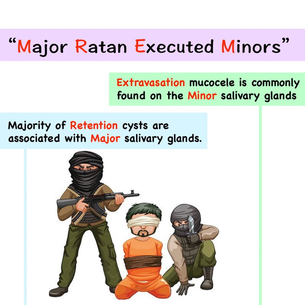
- Extravasation more common than retention cyst.
- Age: more common in children and young adults; with highest frequency in 2nd decade.
- Gender: no gender predilection.
- Site: mucocele can appear at any site of the oral mucosa containing
- salivary glands.
- Most common on lower lip; followed by floor of mouth & ventral tongue > Palate > Buccal mucosa > upper lip; and least common in retromolar pad and posterior dorsal area of the tongue.
Oral Mucocele Clinical features:
- Patients usually complain of a painless swelling which is frequently recurrent.
- Mucoceles can appear at anywhere in the oral mucosa such as lip, cheeks and the floor of the mouth, but mainly appear in the lip. (Ref)
- Duration of lesion may be from few days to months or even years as patients might not seek treatment immediately.
- Swelling may develop suddenly at meal times and many drain spontaneously at intervals.
- Superficial mucocoele may be only 1–2mm in diameter but deeper mucocele is usually larger, the majority being between 5 and 10mm in diameter.
- The swellings are round or oval and smooth.
- Superficial Mucoceles present a bluish, soft and transparent cystic swelling which frequently resolves spontaneously.
- Deeper lesions are the colour of normal mucosa and are firmer.
- Mucoceles associated with the glands of Blandin and Nuhn on the anterior ventralaspect of the tongue are usually less than 10mm in diameter, and usually appear as polypoid or pedunculated swellings: may interfere with normal feeding or breathing.
Pathogenesis of Oral Mucocele:
Two etiological factors in mucoceles: trauma and obstruction of salivary gland ducts
Extravasation mucocele:
- Extravasation mucoceles are caused by a leaking of fluid from surrounding tissue ducts or acini.
- Extravasation mucocele is commonly found on the minor salivary glands.
- Physical trauma can cause a leakage of salivary secretion into surrounding submucosal tissue, stagnant mucous leads to Inflammation.

- Extravasation mucoceles undergo three evolutionary phases.
- First phase: mucous spills diffusely from the excretory duct into conjunctive tissues where some leucocytes and histiocytes are found.
- Second Phase: Granulomas appear during the resorption phase due to histocytes, macrophages and giant multinucleated cells associated with a foreign body reaction.
- Third phase: Connective cells form a pseudocapsule without epithelium around the mucosa.
Retention mucocele:

- Retention mucoceles are formed by dilation of the duct secondary to its obstruction or caused by a sialolith or dense mucosa.
- Majority of retention cysts are associated with major salivary glands.
Histopathology:
- Robinson and Hjørting-Hansen in 1964 described three distinct morphological patterns of mucocoele microscopically.
- The first two represent mucous extravasation cysts and
- Third mucous retention cysts.
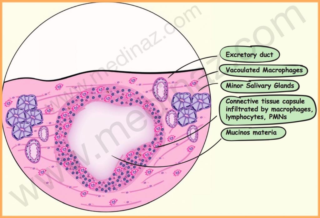
- The cyst contains faintly eosinophilic mucinous material and numerous vacuolated macrophages which are often called ‘muciphages’.
- The mucinous material is surrounded by granulation tissue, or dense fibrous tissue or both.
- The fibrous tissue is infiltrated by vacuolated macrophages, lymphocytes and polymorphonuclear leucocytes, including eosinophils.
- In some cases, a ruptured salivary duct may be identified feeding into the mucinous area. One or more dilated ducts may be present
- Occasionally, mucocele may show epithelial lining, which possibly represents a portion of excretory duct.
- Epithelial lining may also be seen in case of a retention cyst.
- The associated salivary gland, acini which often show alterations:
- May consist of interstitial inflammation or sialadenitis,
- Dilatation of intralobular and interlobular ducts with collection of mucus, and
- Breakdown of individual acinar mucous cells resulting in the formation of tiny areas of pooled mucus
Oral Mucocele Treatment:
- Conventional treatment is surgical extirpation of the surrounding mucosa and glandular tissue down to the muscle layer.
- Some mucoceles are short-lived lesions that rupture and heal by themselves.
- In the case of larger mucoceles, marsupialization may be done to avoid damage to vital structures.
- Whenever possible the associated lobules of salivary gland should be removed with the cyst intact.
Medinaz Dental Cyst Book
“High Yield Visual Book of Dental Cyst” is now available on “Medinaz” App. The App is available on Appstore & Playstore. Visit our website www.medinaz.com for other available books.
Book overview:
– All the necessary High-Yield Points
– 230+ Frequently tested facts
– 200+ hand drawn Images
– Mnemonics to remember
– Helpful for: NBDE, NEET MDS, and Board exams
– FREE UPDATES up to 1 year from the date of publish
– (Time span to be counted from the day it was published)
– Neatly organized materials
– Lifetime access
– Format Image based PDF
A Visual Learning Platform


