
This illustrated notes covered all the important highyield points related to dentigerous cyst
Table of Contents
General features
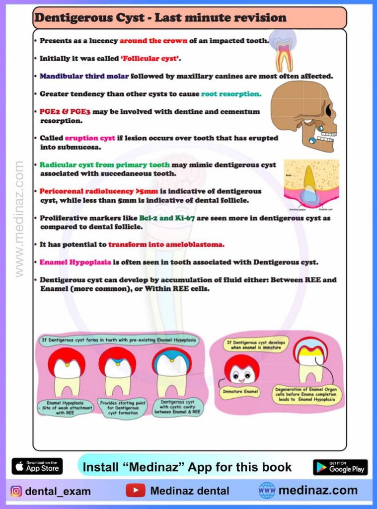
- Dentigerous cyst encloses crown of an unerrupted tooth
- It is attached to CEJ
- Forms by expansion of Dental Follicle
- It was also called Follicular cyst previously
- Brown & Smithe – preferred the term Dentigerous cyst over follicular cyst.
- Dentigerous = Tooth containing
- Follicular = Implies derivation from dental follicle, which is a mesodermal tissue
Epidemiology
- Most common in mandibular 3rd molar followed by maxillary canine > Maxillary 3rd molar > mandibular 2nd premolar
- Peak Occurrence – 3rd decade
- More common in males (1.8:1)
- White > Black (1.6:1)
Clinical features
- Most are incidental finding on a radiograph taken for missing or impacted tooth (failed to erupt)
- Slow enlarging swelling
- Greater tendency than other cysts to cause root resorption
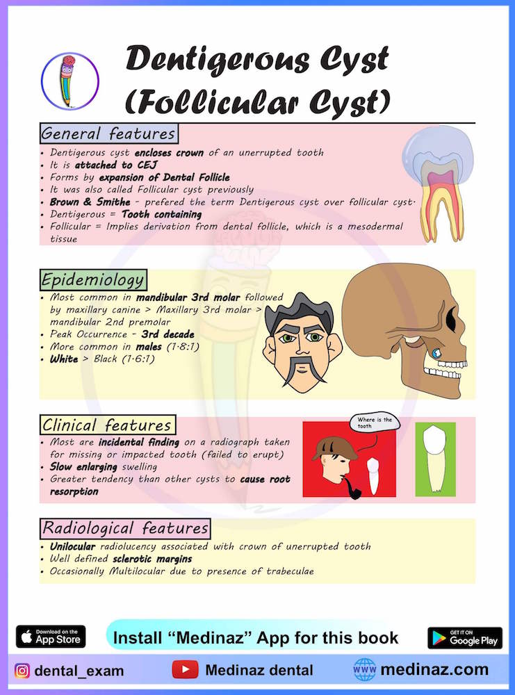
Radiological features of Dentigerous Cyst
- Unilocular radiolucency associated with crown of unerrupted tooth
- Well defined sclerotic margins
- Occasionally Multilocular due to presence of trabeculae
Three radiological variants include:
Central:
Crown is enveloped symmetrically – Tooth often moves in apical direction due to pressure from cystic content. – Mandibular 3rd molar may be pushed towards lower border or into the ramus.
Lateral:
Usually seen in partially erupted teeth. – Dilation of follicle is seen on one side.
Circumferential:
Whole tooth appears to be entrapped by the cyst.
- Radicular cyst from primary tooth may mimic dentigerous cyst associated with succedaneous tooth
- Though it is not common
Distinguishing Dentigerous cyst from dental follicle
- Peri-coronal width (size of cyst) should be at- least 5cm to call it a cyst.
- Less than 5cm should be considered dental follicle.
- Proliferative markers like Bcl-2 & Ki-67 are seen more in dentigerous cyst.
- Apoptotic markers like Fas & ssDNA are more in dental follicle.
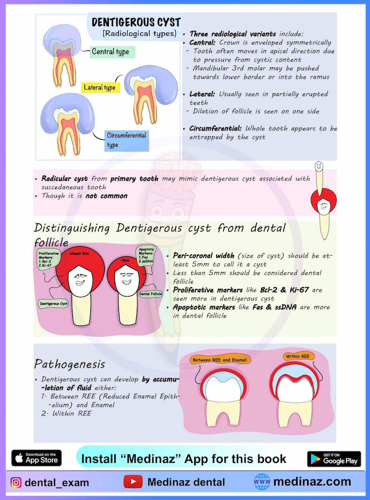
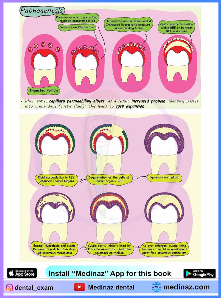
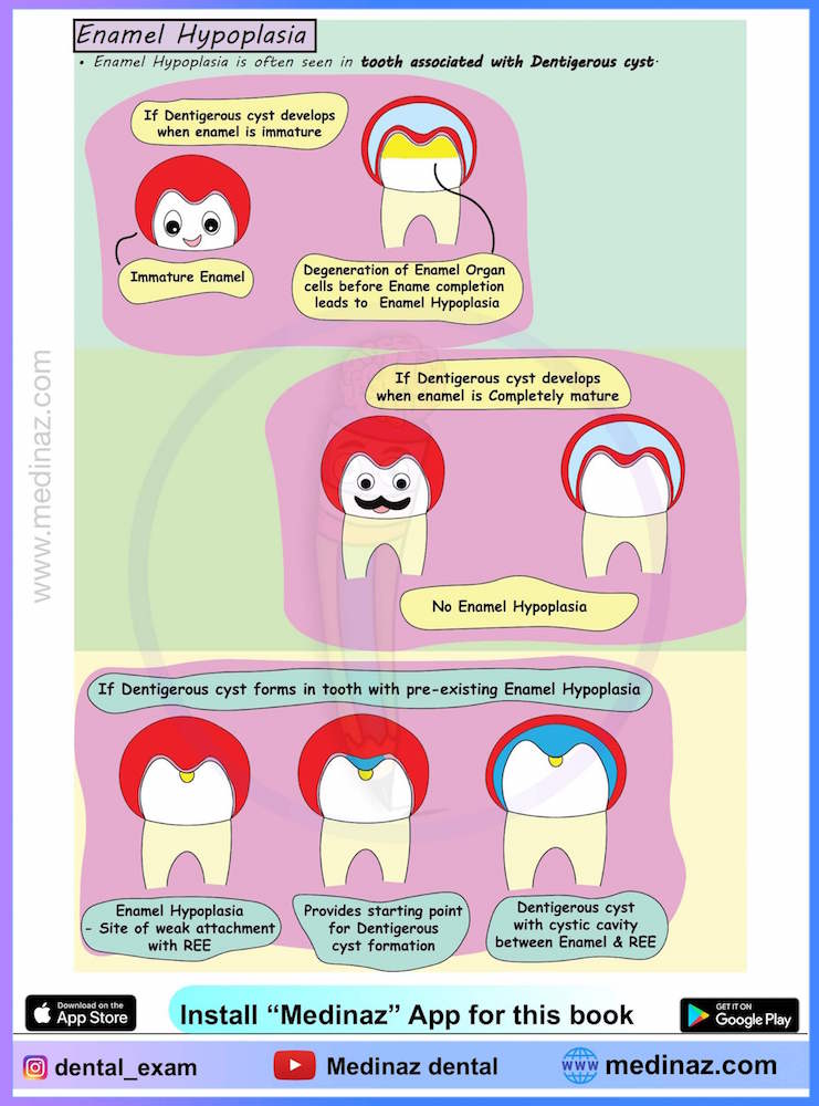
Pathogenesis of Dentigerous Cyst
Histopathology
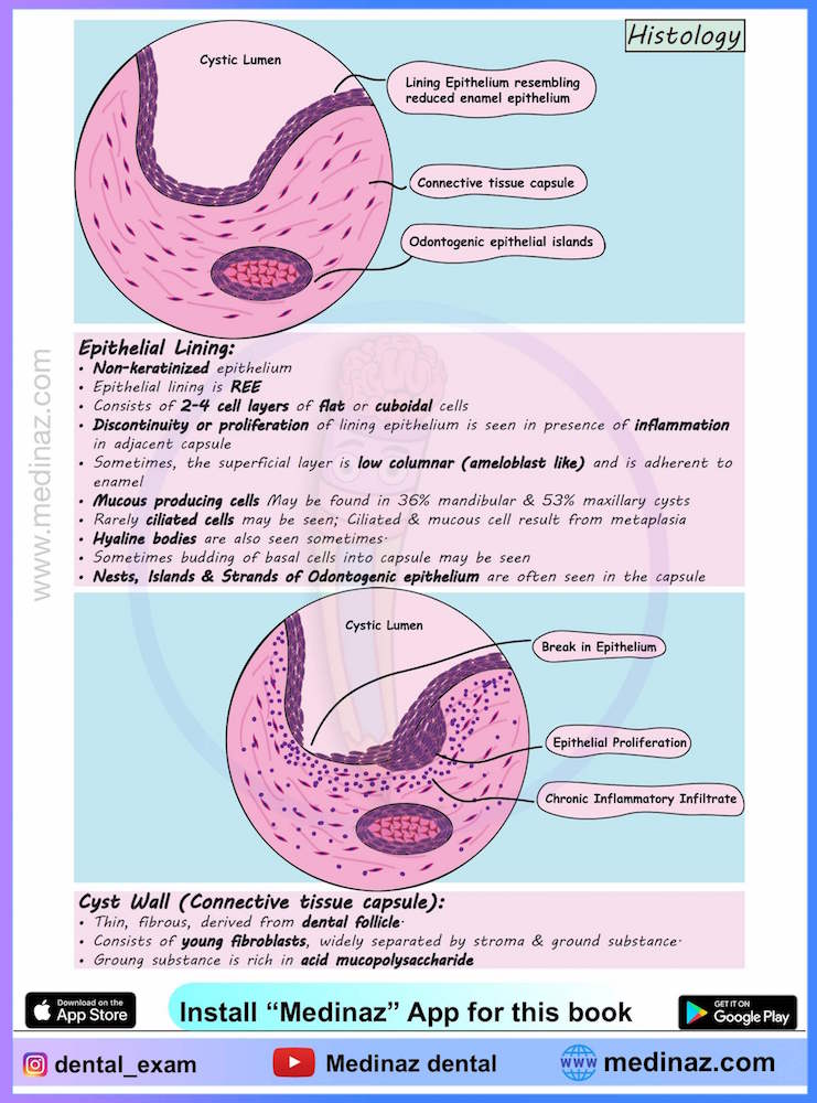
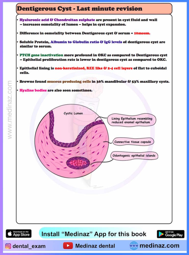
Epithelial lining of dentigerous cyst
- Non-keratinized epithelium
- Epithelial lining is REE
- Consists of 2-4 cell layers of flat or cuboidal cells
- Discontinuity or proliferation of lining epithelium is seen in presence of inflammation in adjacent capsule
- Sometimes, the superficial layer is low columnar (ameloblast like) and is adherent to enamel
- Mucous producing cells may be found in 36% mandibular & 53% maxillary cysts
- Rarely ciliated cells may be seen; Ciliated & mucous cell result from metaplasia
- Hyaline bodies are also seen sometimes
- Sometimes budding of basal cells into capsule may be seen
- Nests, Islands & Strands of Odontogenic epithelium are often seen in the capsule.
Cyst Wall (Connective tissue capsule)
- Thin, fibrous, derived from dental follicle
- Consists of young fibroblasts, widely separated by stroma & ground substance
- Groung substance is rich in acid mucopolysaccharide.
Treatment
- Emphasis is given on conservative surgical treatment with orthodontics to retain the involved tooth & ensure its eruption into normal occlusion.
- Marsupialization may also be employed
- Teeth with incomplete root formation have better chance to erupt, as eruption is closely related to root development.
Last minute revision
Medinaz Dental Books
“High Yield Visual Book of Dental Cyst” is now available on “Medinaz” App. The App is available on Appstore & Playstore. Visit our website www.medinaz.com for other available books.
Book overview:
– All the necessary High-Yield Points
– 230+ Frequently tested facts
– 200+ hand drawn Images
– Mnemonics to remember
– Helpful for: NBDE, NEET MDS, and Board exams
– FREE UPDATES up to 1 year from the date of publish
– (Time span to be counted from the day it was published)
– Neatly organized materials
– Lifetime access
– Format Image based PDF
A Visual Learning Platform


