
Benign Cementoblastoma Radiology: Dental Notes for BDS, MDS, NBDE Exams.
Synonyms
- Cementoblastoma
- True cementoma
Disease Mechanism
- Slow-growing mesenchymal neoplasm composed of cementum-like tissue.
- Manifests as a bulbous growth around and attached to the apex of a tooth root.
- Histologic characteristics are identical to osteoblastomas.
- Some authors classify cementoblastomas as bone tumors due to these similarities.
- Typically develops with permanent teeth; rare cases involve primary teeth.
Clinical Features
- Uncommon lesion but may be more prevalent than reported.
- More common in males than females.
- Age range: 12–65 years; most patients are relatively young.
- No racial predilection.
- Usually a solitary, slow-growing lesion that may eventually displace teeth.
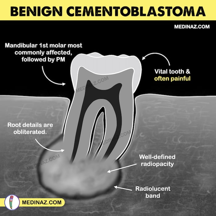
- The involved tooth is vital and often painful.
- Pain varies among patients; can be relieved by anti-inflammatory drugs.
Imaging Features
Location
- Predominantly occurs in the mandible (78%).
- Commonly forms on the roots of a premolar or first molar (90%).
Periphery
- Appears as a well-defined radiopacity with a cortical border.
- Surrounded by a well-defined radiolucent band just inside the cortical border.
Internal Structure
- Mixed radiolucent-radiopaque lesion; predominantly radiopaque internally.
- May display an amorphous pattern or a wheel-spoke pattern.
- Cemental mass density often obscures the root outline.
- Central radiopaque mass surrounded by a radiolucent band, indicating maturation from center to periphery.
Effects on Surrounding Structures
- External root resorption may be visible if the root outline is apparent.
- Large tumors can cause mandibular expansion.
- Possible perforation through the outer cortical plate without periosteal reaction.
Differential Diagnosis
Periapical Osseous Dysplasia
- Radiolucent band is usually less defined and uniform compared to cementoblastoma.
- Has a more irregular, undulating outline.
Periapical Sclerosing Osteitis
- Lacks a soft tissue capsule present in cementoblastomas.
Dense Bone Island (DBI)
- Also lacks a soft tissue capsule.
Hypercementosis
- Surrounded by a periodontal membrane space, which is thinner than the cementoblastoma’s capsule.
- Does not cause root resorption or jaw expansion.
Treatment
- Self-limiting; rarely recurs after enucleation.
- Simple excision and extraction of the associated tooth are usually sufficient.
- The gold-standard treatment: total curettage and surgical excision of the lesion followed by extraction of affected tooth structures. (Ref)
- Tumor may be amputated from the tooth in some cases, followed by endodontic treatment.
“Visual Book of Odontogenic Tumor” is now available on “Medinaz” App. The App is available on Appstore & Playstore. Visit our website www.medinaz.com for other available books.
Book overview:
– All the necessary High-Yield Points
– 550+ Frequently tested facts
– 300+ hand drawn Images
– Mnemonics to remember
– Helpful for: NBDE, NEET MDS, and Board exams
– FREE UPDATES up to 1 year from the date of publish
– (Time span to be counted from the day it was published)
– Neatly organized materials
– Lifetime access
– Format Image based PDF
Benign Cementoblastoma Radiology Reference:
- Oral radiology Principles and Interpretation; White & Pharoah
- Essentials of Oral and Maxillofacial Radiology; F. Karjodkar
- Concise Oral Radiology; HR Umarji
A Visual Learning Platform


