
Fibrous Dysplasia Radiology – Location, periphery, internal structure, effects on surrounding structures
Table of Contents
1. Location
Involves the maxilla almost twice often as the mandible
Most commonly posterior region
Commonly unilateral and very rarely bilateral
2. Periphery
Periphery of the lesion is most commonly ill-defined and blends imperceptibly with the normal bone
The periphery of the young lesions sometimes appears to be sharp and even corticated.
3. Internal structures
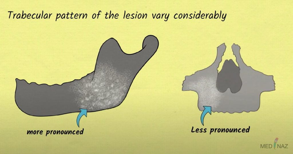
Density and trabecular pattern of the lesion vary considerably
Variation is more pronounced in Mandible and more homogenous in maxilla
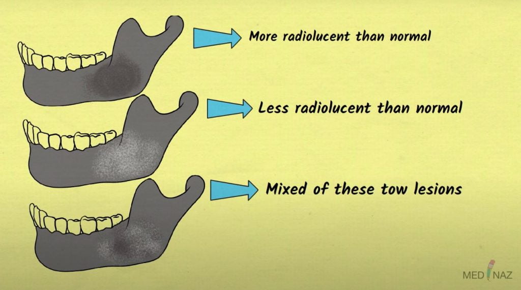
The internal aspect of bone is of 3 types
- More radiolucent than normal
- Less radiolucent than normal
- Mixture of these 2 lesions
The early lesions appear as a cyst like radiolucency in the jaws
Sometimes it appears to have granular internal septa, giving internal aspect a multilocular appearance
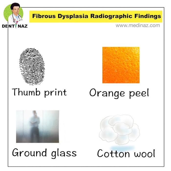
Trabeculae – Abnormal trabeculae usually are shorter, thinner, irregularly shaped and more numerous than normal trabeculae
The altered trabeculae may give rise to various appearance such as-
-Orange peel (Peau D’orange)
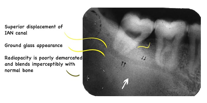
-Ground glass
-Thumb print
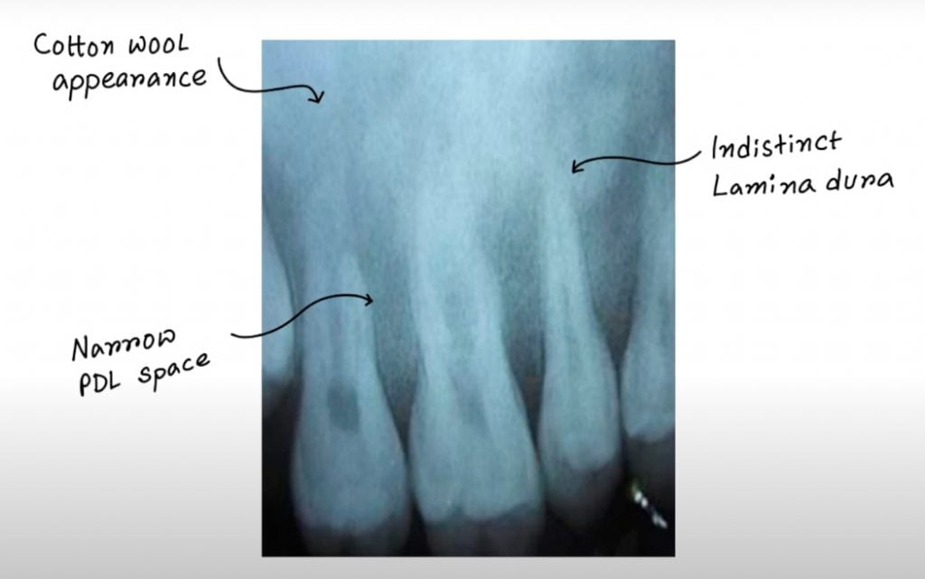
-Cotton wool
Simple bone cyst like bone cavities are seen and occurs more commonly in mandibular lesions
4. Effects on surrounding structures
Small lesion has no effect on surrounding structure
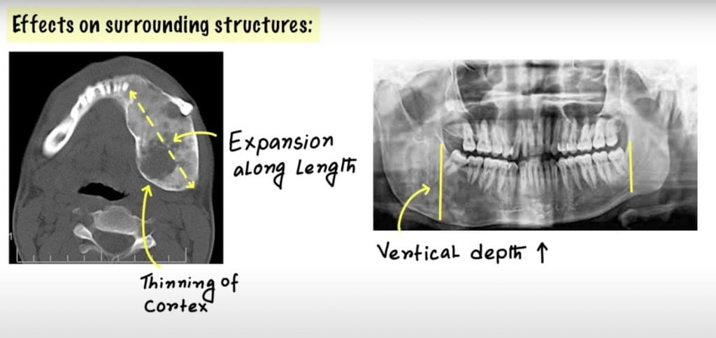
Typically cause enlargement of the bone from within cause ribbon like thinning of cortex
Expansion of the bone is even along it’s length rather than the more concentric expansion seen with benign tumors
Vertical depth of the mandible is often increased
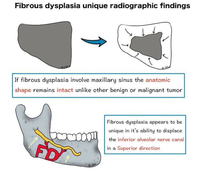
In the maxilla the lesion encroaches the sinus usually from the lateral-wall and last section of the sinus to be involved is the most postero-superior portion
Normal anatomic shape of the antrum is most oftenly maintained
Lamina dura of the teeth in the affected area of the bone become indistinct.
PDL space may appear to be very narrow
FD can displace teeth and interfere with normal eruption
Superior displacement of the IAN can is another typical finding of FD Rarely there is root resorption
A Visual Learning Platform


